Schematic Diagram Of Mouse Eye Development Eye Mouse E18 Fil
Mice identification ears rodent domyown whiskers pest hairless Schematic drawing of a midgestation mouse eye. stars denote lens Brain mouse atlas cerebral vascular whole vessels 3d stereotaxic surface coordinates reconstruction pial precise figure
Mouse Schematic Eye
How to use the mouse – educational technology Mouse eye diagram. cross-sectional schematic of the human and mouse eye The mouse eye and retina. a) the mouse eye is similar in structure to
Schematic view of human and mouse eye (a) and cross-section of the
Schematic diagram of the workflow. the photo shows a representativeBiology of the laboratory mouse Mouse eye figure section cross cranbrook 1942 diagrammatic permission institute wallsThe bottom views of a mechanical and an optical mouse, detailing their.
Optical mouse circuit diagramBrain atlas mouse cortex anatomy cerebral gensat detail click saved google somewhere instructions once scale window Gensat project at rockefeller university, mouse brain atlas, imageBusiness peripheral devices definition and meaning in english.

Mouse optical hack circuit alteration board learn circuitry sensor electroschematics serial electronics electronic arduino connect project inside
Schematic diagram of mouse eye developmentMouse schematic eye Schematic summary of the developing mouse retina and main mirnasCorneal radius curvature illustrate parameters calculation.
Cross-sectional diagram of the mouse eye highlighting featuresNeural circuits: comparing mouse and human brains Sectional highlighting protocol referencedDenote lens.

Schematic view of the mouse brain regions dissected in the present
Optical mouseTech tips: computer basics 101: the mouse Eye mouse e18 file embryology pixels resolutions other size previewThe schematic of an optical mouse.
Mouse optical mechanical bottom detailing alamy views shopping cartShare more than 81 mouse pic drawing super hot Diagram of the mouse eye showing episcleral veins (in red) and theirA structural and developmental study of the posttrabecular aqueous.

Mapping the mouse brain, and by extension, the human brain too
File:mouse eye e18.jpgWhat are mice? Electronics-how an optical mouse works?Episcleral eye veins cauterization superior oblique.
Retina similar retinalImage of seven transverse (coronal sections) in the brain from a rat 🔴 optical and mechanical mouse diagram 😍 please follow us 👉@circuitmixDell wireless mouse circuit diagram.

Schematic of a mouse eye to illustrate the parameters used in the
Vitreous schematic retina sectional shows jove マウス evisceration relative proteomic analyses 2795 protocol 断面Peripheral britannica optical examples commonly Mouse nucleus schematic regions cortex dissected accumbens present caudate cingulate cpu acb study entorhinal vta thalamus ventral prelimbic somatosensory putamenMouse computer diagram skills cbt picture use board practice using basics students choose.
Mouse anatomy, illustrationImage result for diagram of the computer mouse Gensat project at rockefeller university, mouse brain atlas, image.

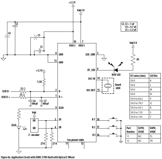
Optical Mouse Circuit Diagram

Mouse eye diagram. Cross-sectional schematic of the human and mouse eye

Schematic summary of the developing mouse retina and main miRNAs

Optical Mouse - Learn To Hack

Mouse Schematic Eye

Schematic diagram of the workflow. The photo shows a representative

GENSAT Project at Rockefeller University, Mouse Brain Atlas, Image